Osteonecrosis (Avascular Necrosis) In Singapore

Osteonecrosis, also known as avascular necrosis (AVN), is a medical condition where bone tissue dies due to a disrupted blood supply. When it affects the hip joint, it can cause pain and disability if not properly managed. With early detection and the right treatment, it is possible to protect the joint and reduce the risk of future complications.

doctor img
Dr Zackary Chua

MBBS (Aus) MMed (SG) MRCS (Glasgow) FRCSEd (Orth)

Osteonecrosis w alpha Osteonecrosis (Avascular Necrosis)

What is Osteonecrosis?

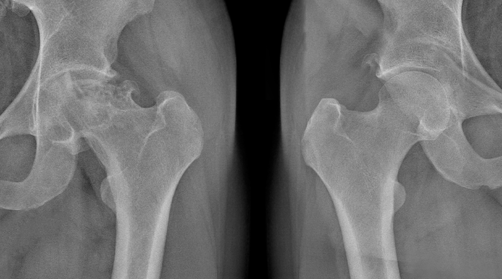
Osteonecrosis happens when blood flow to a part of a bone is disrupted, resulting in the death of the bone tissue. In the hip, this affects the femoral head (the ball portion of the ball-and-socket joint). Insufficient blood supply causes the bone to weaken, collapse, and may eventually lead to arthritis and joint damage.

Types of Osteonecrosis

Traumatic Osteonecrosis

Occurs from physical injury to the hip, such as hip fractures or dislocations. The trauma can damage blood vessels supplying the femoral head, leading to immediate or delayed bone death. Symptoms may appear within months to two years after the initial injury.

Non-traumatic Osteonecrosis

It develops without obvious injury. Various systemic conditions and risk factors gradually compromise blood flow to the hip joint over time. Non-traumatic osteonecrosis often affects both hips, with bilateral involvement occurring in many cases.

Idiopathic Osteonecrosis

When no clear cause can be identified, the condition is classified as idiopathic. This may involve genetic predisposition or unrecognised risk factors.

Causes & Risk Factors

Direct Causes

  • Hip fractures, particularly femoral neck fractures
  • Hip dislocations that damage blood vessel
  • Radiation therapy to the pelvic region
  • Decompression sickness (“the bends”) in divers
  • Blood disorders such as sickle cell disease

Major Risk Factors

  • Long-term corticosteroid use (the most common risk factor)
  • Excessive alcohol consumption (more than three drinks per day)
  • Smoking
  • Systemic lupus erythematosus (SLE)
  • Organ transplantation requiring immunosuppression
  • HIV/AIDS and related treatments
  • Gaucher’s disease
  • Pancreatitis
  • Hyperlipidaemia (high cholesterol and triglycerides)
  • Chemotherapy

Signs & Symptoms

Early Stage

  • Mild groin pain during weight-bearing activities
  • Occasional stiffness in the hip
  • Discomfort that improves with rest
  • No visible changes on standard X-rays

Advanced Stage

  • Severe groin pain, even at rest
  • Limited hip range of motion
  • Limping or altered gait
  • Pain radiating to knee or buttock
  • Difficulty climbing stairs or rising from seated position
  • Night-time pain disrupting sleep

Late Stage

  • Constant, debilitating hip pain
  • Noticable leg length discrepancy
  • Muscle weakness and atrophy
  • Complete loss of hip function
  • Need for walking aids
  • Visible hip deformity

Symptoms develop gradually over months to years. The progression rate varies between individuals, with some experiencing rapid deterioration while others maintain stable symptoms for extended periods.

Don’t wait until hip damage worsens.

Schedule a consultation to understand your condition, review potential management strategies, and learn how timely intervention can support long-term joint health.

When to See a Doctor

Seek medical attention if you experience persistent groin or hip pain lasting more than a few days, especially if you have known risk factors for osteonecrosis. Urgent evaluation is required for red-flag symptoms, including sudden severe hip pain after minor trauma, inability to bear weight, or hip pain accompanied by fever or other signs of infection.

Early consultation is important, as treatment outcomes are closely linked to the stage at diagnosis. During your first visit, our orthopaedic surgeon will conduct a thorough medical history review, preform a physical examination, and order appropriate imaging studies. The consultation involves discussing your symptoms, risk factors, and impact on daily activities. Based on findings, a personalised treatment plan will be developed to address your specific condition stage and lifestyle needs.

Diagnosis & Testing Methods

Accurate diagnosis requires clinical evaluation and imaging. Standard X-rays may appear normal in early stages, but as the condition progresses, they can reveal characteristic changes such as crescent signs, femoral head flattening, and joint space narrowing.

Magnetic Resonance Imaging (MRI) is the preferred method for early detection, as it can identify bone marrow changes before structural damage occurs. Computed Tomography (CT) scans provide detailed views of bone architecture, which is particularly useful for surgical planning. Bone scans using radioactive tracers can also identify areas of increased bone activity.

Laboratory tests help identify underlying causes and risk factors. These include complete blood count, inflammatory markers, lipid profiles, and specific tests for autoimmune conditions. Laboratory results are typically available within 3–5 days, while MRI reports are usually ready within 24–48 hours, allowing timely assessment and management planning.

Treatment Options Overview

Conservative Management

Physiotherapy helps maintain hip function and strength. Exercise programmes focus on range of motion, muscle strengthening, and gait training while avoiding excessive joint stress. Aquatic therapy offers low-impact conditioning, which can be particularly beneficial for symptomatic patients.

Medications

Non-steroidal anti-inflammatory drugs (NSAIDs) help manage pain and inflammation. For patients with gastrointestinal concerns, selective COX-2 inhibitors are an alternative. Bisphosphonates may be considered to slow bone resorption and disease progression, though evidence remains mixed regarding long-term efficacy.

Activity Modification

Using walking aids or crutches reduces weight-bearing stress on the affected hip. Lifestyle adjustments include avoiding high-impact activities, maintaining a healthy body weight, and modifying daily or work environments to minimise hip strain.

Core Decompression

This minimally invasive procedure involves drilling small channels into the femoral head to reduce intraosseous pressure and stimulate new blood vessel formation.

Bone Grafting Procedures

Vascularised fibular grafts provide structural support and blood supply to the necrotic area. Non-vascularised grafts combined with growth factors offer an alternative for smaller lesions. These procedures aim to prevent femoral head collapse and delay joint replacement need.

Osteotomy

Rotational osteotomies reposition healthy bone to the weight-bearing surface. This technically demanding procedure suits younger patients with localised disease.

Hip Resurfacing

Metal-on-metal resurfacing preserves more bone than total hip replacement. Suitable for younger, active patients with good bone quality.

Total Hip Replacement

When joint preservation fails, total hip arthroplasty provides reliable pain relief and function restoration. Different implant types, such as ceramic-on-ceramic or ceramic-on-polyethylene, can be considered based on patient-specific anatomy and activity needs.

Persistent hip or groin pain can worsen if left unassessed.

Explore non-surgical and surgical options, and learn which treatment plan is most suitable for your condition.

Complications if Left Untreated

If osteonecrosis is not addressed, the affected bone may collapse, leading to severe arthritis in the hip joint. Most untreated cases progress to significant joint damage within 2–3 years of symptom onset. The resulting irregular joint surfaces accelerate cartilage wear, causing painful bone-on-bone contact.

Chronic pain and disability can affect quality of life. Patients often develop compensatory movement patterns, which may lead to back pain, problems in the opposite hip, or knee strain. Advanced disease can make basic activities such as walking, dressing, or climbing stairs difficult, sometimes necessitating walking aids or wheelchair use.

Long-term complications include chronic pain syndrome, muscle atrophy, osteoporosis from disuse, and increased fall risk.

Prevention

While not all cases of osteonecrosis can be prevented, certain strategies can help reduce risk. Limit corticosteroid use to the lowest effective dose, moderate alcohol intake, and stop smoking to support healthy blood flow. Managing underlying conditions such as lupus, sickle cell disease, and high cholesterol is also important.

For high-risk individuals, regular monitoring allows early detection. This includes periodic hip examinations and MRI screening for those on long-term steroids or with bilateral risk factors. Maintaining healthy body weight reduces mechanical stress on hip joints.

Osteonecrosis (Avascular Necrosis)

Frequently Asked Questions

Can osteonecrosis of the hip heal on its own?

Established osteonecrosis rarely heals spontaneously. Very Small, early-stage lesions may sometimes stabilise, but most cases progress due to the disrupted blood supply, which prevents natural bone repair.

How long does recovery take after hip preservation surgery?

Recovery varies by procedure and individual factors. Core decompressiontypically requires 6-12 weeks of crutch use, with a full return to activities in 3-4 months. Vascularised bone grafts often need 3–4 months of limited weight-bearing, while osteotomy procedures may require 3–6 months for bone healing. Physiotherapy continues throughout recovery to restore strength and flexibility, with complete improvement potentially taking up to one year.

Will I need hip replacement eventually even with early treatment?

Not necessarily. Early-stage, pre-collapse osteonecrosis can often be managed with joint preservation procedures, depending on lesion size, location, age, and management of risk factors. Larger lesions or post-collapse cases may progress despite treatment, so ongoing monitoring is important to determine if and when hip replacement becomes necessary.

Can osteonecrosis affect both hips simultaneously?

Yes, bilateral involvement is common, especially in non-traumatic cases. Risk factors such as long-term steroid use, alcohol consumption, and certain systemic conditions can affect both hips. Screening the opposite hip with MRI is recommended even if there are no symptoms, allowing early detection and informed treatment planning.

What activities should I avoid with hip osteonecrosis?

Activity restrictions depend on disease stage and treatment type. Avoid high-impact activities like running, jumping, and contact sports. Repetitive impact activities such as jogging on hard surfaces should be replaced with swimming, cycling, or elliptical training. Heavy lifting and prolonged standing may also stress the hip joint unnecessarily. Our orthopaedic surgeon will provide specific guidelines based on your condition severity and treatment plan. The goal is maintaining fitness while protecting the hip from excessive stress.

Is osteonecrosis hereditary?

While osteonecrosis itself isn’t directly inherited, certain genetic factors increase susceptibility. Inherited conditions affecting blood clotting (thrombophilia), fat metabolism (hyperlipidaemia), or bone metabolism may predispose to osteonecrosis. Genetic variants affecting alcohol and steroid metabolism also influence risk. Family history of hip problems warrants discussion during consultation. Most cases result from acquired risk factors rather than genetic predisposition alone.

Conclusion

Osteonecrosis of the hip is a serious condition that benefits from early detection and timely intervention. Recognising symptoms early allows for appropriate management before significant joint damage occurs. Treatment options range from joint-preserving procedures in the initial stages to reconstructive approaches for more advanced cases. A comprehensive, personalised approach helps address current symptoms while supporting long-term hip function and mobility.

If you have ongoing hip discomfort, don’t wait.

Schedule a consultation to assess the cause and explore management options tailored to you.

Dr Zackary Chua 1

Dr Zackary Chua (蔡克祥医生)

MBBS (AUS)|MMed (Singapore)|MRCS (Glasgow)|FRCSEd (Orth)

A senior consultant orthopaedic surgeon with dual specialisations in Foot & Ankle Surgery and Paediatric Orthopaedics. Backed by his extensive years of training and experience, Dr Chua brings the necessary knowledge and skills to ensure a smooth recovery journey.

Rooted firmly by two fundamental philosophies: “When one’s feet hurt, one hurts all over” and “Children are not little adults”, Dr Chua’s approach to patient care reflects his compassionate care for adults and children.

  • Singhealth Quality Excellence Award – Silver (2011)
  • Service From the Heart Award (2021)
  • Singapore Health Quality Service Award – Gold (2023)
  • NUS Medicine Dean’s Award for Teaching Excellence (2023)

His active participation in the medical community extends to mentoring, research supervision, and contributions to key professional forums and symposiums, both locally and internationally.

Your Treatment Roadmap

Registration

Our friendly clinical staff will assist you with your registration, ensuring an efficient and hassle-free process for you.

Specialist Consultation

During your consultation, we will evaluate your medical history and the pain you are experiencing. You may also ask questions about your condition.

Diagnosis Of Your Condition

We will likely perform some form of diagnostic imaging (e.g. X-Rays, MRI) to assess your condition accurately.

individualised Treatment Plan

After imaging, we will review the results of your scans thoroughly, and advise an individualised treatment plan for you.

Follow-Up Visits

As you go through your treatment plan, we will follow up with you every step of the way, ensuring that you receive the dedicated care you deserve.

Billing & Insurance

Integrated Shield Plans & Corporate Insurance

For Singaporeans, Singapore Permanent Residents and Foreigners.
Please speak to our friendly clinic staff about using your insurance plans.

Make An Enquiry

Got a Question? Fill up the form and we will get back to you shortly.

    Full Name*

    Email Address*

    Phone Number*

    Your Message*

    For Faster Response, Call us!

    +65‎ 6956‎ 6588


    Our Locations

    Mount Elizabeth Novena Specialist Centre
    38 Irrawaddy Road, #05-36, Singapore 329563

    +65‎ 6956‎ 6588 (24 hours) (Calls / Messages)
    +65 8877 3318 (24 hours) (WhatsApp)
    Monday – Friday: 9am – 5pm
    Saturday: 9am – 12:30pm
    Sunday & PH: Closed

    Parkway East Medical Centre
    319 Joo Chiat Place, #03-06, Singapore 427989

    +65‎ 6956‎ 6588 (Calls / Messages)
    +65 8877 3318 (WhatsApp)
    Monday – Friday: 9am – 5pm
    Saturday: 9am – 12:30pm
    Sunday & PH: Closed
    (Parkway East Clinic is on appointment basis)