Articular Cartilage Injury

Knee pain from an articular cartilage injury can limit mobility and affect quality of life. Whether you’re an athlete experiencing persistent joint discomfort or someone experiencing knee pain after an injury, understanding cartilage damage is the first step toward effectivecare. At our specialist clinic in Singapore, we offer thorough assessment and evidence-based treatment options for articular cartilage injuries to help patients maintain joint health and function.

doctor img
Dr Zackary Chua

MBBS (Aus) MMed (SG) MRCS (Glasgow) FRCSEd (Orth)

Articular cartilage injury Articular Cartilage Injury

What is Articular Cartilage Injury?

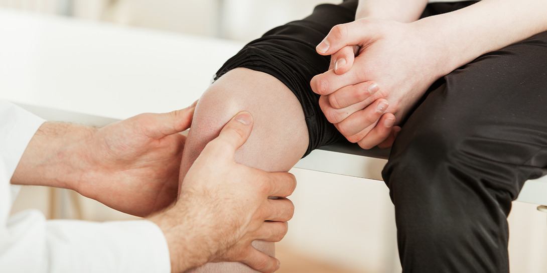
Articular cartilage is the smooth, white tissue covering the ends of bones where they meet to form joints. In the knee, this specialised tissue allows bones to glide smoothly against each other with minimal friction. An articular cartilage injury occurs when this protective layer becomes damaged through trauma, wear, or degenerative processes.

Unlike other tissues in the body, articular cartilage has a limited blood supply and poor self-healing capacity. Once damaged, it rarely heals completely on its own. Studies show that cartilage injuries affect approximately 15-20% of adults in Singapore, with higher prevalence among active individuals and those over 40 years old.

Types of Articular Cartilage Injury

Acute Traumatic Injuries

These injuries result from sudden impact or twisting movements during sports or accidents. The cartilage may crack, tear, or separate from the underlying bone. Common in younger athletes, these injuries often occur alongside other knee damage, such as ligament tears.

Degenerative Cartilage Damage

This type develops gradually due to repetitive stress and ageing. The cartilage slowly wears away, becoming thinner and rougher. This process typically begins after age 40 and may progress to osteoarthritis if left untreated.

Osteochondral Lesions

These injuries involve both the cartilage and the underlying bone. A piece of cartilage, along with attached bone, may partially or completely detach from the joint surface. These lesions can occur from acute trauma or develop through repetitive microtrauma.

Chondral Flaps and Loose Bodies

Damaged cartilage creates flaps that catch during knee movement or break off entirely, forming loose fragments within the joint. These loose bodies can cause locking, catching, and additional damage to healthy cartilage surfaces.

Causes & Risk Factors

Causes

  • Direct trauma from falls, collisions, or sports injuries
  • Sudden twisting or pivoting movements, placing excessive stress on the knee
  • Repetitive impact from high-impact activities over many years
  • Previous knee surgeries or injuries weakening the joint structure
  • Underlying conditions affecting cartilage health, such as osteochondritis dissecans

Risk Factors

  • Age over 40 years, as cartilage naturally becomes less resilient
  • Participation in high-impact sports (basketball, football, running)
  • Previous knee injuries, particularly ACL tears or meniscus damage
  • Excess body weight increases stress on knee joints
  • Genetic predisposition to cartilage problems
  • Occupations requiring frequent kneeling, squatting, or heavy lifting
  • Joint malalignment, placing uneven pressure on cartilage surfaces

Signs & Symptoms

Mild Symptoms

  • Intermittent knee pain during or after activity
  • Mild swelling after prolonged use
  • Slight stiffness in the morning lasting less than 30 minutes
  • Occasional clicking or grinding sensation
  • Minor discomfort when climbing stairs

Moderate Symptoms

  • Persistent pain even with routine daily activities
  • Noticeable swelling lasting several days
  • Difficulty fully bending or straightening the knee
  • Feeling of instability or giving way
  • Pain that wakes you at night
  • Need to modify activities due to discomfort

Severe Symptoms

  • Constant pain significantly limiting mobility
  • Knee locking or catching prevents movement
  • Visible deformity or significant swelling
  • Unable to bear weight on the affected leg
  • Complete inability to participate in sports or exercise

Symptoms typically develop gradually for degenerative injuries but appear suddenly with acute trauma. The severity often correlates with the size and location of the cartilage damage.

Experiencing these symptoms? Don’t wait for them to worsen.

Schedule a consultation with our Senior Consultant Orthopaedic Surgeon for an accurate diagnosis and personalised treatment plan.

When to See a Doctor

If you experience sudden severe pain, significant swelling, or inability to bear weight after a knee injury, please seek immediate medical attention if you. These symptoms may indicate substantial cartilage damage requiring prompt evaluation.

If you experience persistent knee pain lasting more than two weeks, recurring swelling, or mechanical symptoms such as locking or catching, consider scheduling a specialist consultation. Early evaluation can help identify cartilage damage and guide appropriate management. Prompt assessment may also reduce the risk of further joint deterioration.

During your first consultation, our orthopaedic surgeon will perform a comprehensive knee examination, review your medical history, and discuss your symptoms and activity goals. This thorough assessment helps determine the extent of cartilage damage and guides treatment planning.

Diagnosis & Testing Methods

  • Clinical Examination: Our orthopaedic surgeon begins with a detailed knee examination, assessing range of motion, stability and areas of tenderness. Imaging such as X-rays or MRI may be arranged to provide a clearer picture of cartilage health and guide management planning.
  • X-rays: While cartilage doesn’t appear on X-rays, these images show joint space narrowing, bone changes, and alignment issues. X-rays help exclude fractures and assess overall joint health.
  • MRI Scanning: Magnetic resonance imaging provides detailed images of soft tissues including cartilage. This non-invasive scan reveals the location, size, and depth of cartilage lesions. High-resolution MRI can detect even small areas of damage.
  • Diagnostic Arthroscopy: When MRI results are inconclusive, arthroscopy allows direct visualisation of cartilage surfaces. A small camera inserted through tiny incisions provides clear images of the extent of damage. This procedure can transition from diagnostic to therapeutic if treatable lesions are found. Results from imaging studies typically become available within 24-48 hours. Our team will schedule a follow-up appointment to discuss findings and treatment recommendations based on your specific cartilage injury pattern.

Treatment Options Overview

Conservative Management

Initial treatment focuses on reducing symptoms and preventing further damage. This includes activity modification, avoiding high-impact activities while maintaining joint motion through low-impact exercises. Weight management reduces stress on damaged cartilage, with every kilogram of weight loss resulting in four kilograms less pressure on the knee join

Injection Therapies

Corticosteroid injections provide rapid anti-inflammatory effects, typically offering relief for 3-6 months. These injections work well for managing acute flare-ups but aren’t suitable for long-term use due to potential cartilage effects.

Viscosupplementation

Hyaluronic acid injections supplement the knee’s natural joint fluid, improving lubrication and potentially providing protective effects. Treatment involves a series of 3-5 weekly injections, with benefits lasting 6-12 months in responsive patients.

Corticosteroid Injections

When discomfort persists, corticosteroid injections can be considered to help reduce inflammation and fluid production. They may be used alone or after aspiration. More than one injection may sometimes be required.

Viscosupplementation

Hyaluronic acid injections supplement the knee’s natural joint fluid, improving lubrication and potentially providing protective effects. Treatment involves a series of 3-5 weekly injections, with benefits lasting 6-12 months in responsive patients.

Platelet-Rich Plasma (PRP)

PRP therapy uses concentrated platelets from your own blood to promote healing. The growth factors in PRP may stimulate cartilage repair and reduce inflammation. This treatment shows promise for early-stage cartilage injuries, though multiple injections may be needed.

Arthroscopic Procedures

Arthroscopic debridement removes loose cartilage fragments and smooths damaged surfaces, reducing mechanical symptoms. While this doesn’t restore cartilage, it can significantly improve function and comfort for appropriately selected patients.

Microfracture Technique

This arthroscopic procedure creates small holes in the bone beneath damaged cartilage, stimulating bleeding and formation of repair tissue. Microfracture can achieve favourable results in selected patients according to published studies.

Osteochondral Autograft Transplantation (OATS)

OATS involves transferring healthy cartilage and bone from a non-weight-bearing area to the damaged site. This technique works well for medium-sized defects in active patients, providing immediate structural repair with the patient’s own tissue.

Autologous Chondrocyte Implantation (ACI)

ACI represents modern cartilage restoration, harvesting the patient’s own cartilage cells, growing them in a laboratory, then reimplanting them into the defect. This two-stage procedure suits larger defects in younger patients committed to extensive rehabilitation.

Osteotomy

For patients with cartilage damage and malalignment, osteotomy reshapes the bone to redistribute weight away from damaged areas. This joint-preserving surgery can delay or prevent the need for knee replacement in selected cases.

Partial Knee Replacement

When cartilage damage is limited to one compartment of the knee, partial replacement preserves healthy tissue while resurfacing only the damaged area. This option may offer pain relief while maintaining more natural knee function than total replacement.

Every patient’s condition is unique.

Our Senior Consultant Orthopaedic Surgeon will assess your specific situation and recommend the most suitable treatment approach for you.

Complications if Left Untreated

Without treatment, articular cartilage injuries may progress over time. Damaged cartilage has limited healing capacity and continued use can worsen deterioration. Studies show that as cartilage loss progresses, joint space narrows and bones may develop reactive changes such as osteophytes (bone spurs). These changes alter joint mechanics, increasing stress on remaining healthy cartilage and potentially accelerating degeneration.

Prevention

While not all cartilage injuries are preventable, several strategies reduce risk. Maintaining a healthy weight minimises chronic stress on knee cartilage. Regular low-impact exercise like swimming or cycling strengthens supporting muscles without excessive joint loading.

Proper sports technique and appropriate equipment help prevent acute injuries. Gradual training progression allows tissues to adapt to increasing demands. Cross-training varies stress patterns, reducing repetitive strain on cartilage surfaces.

For those with previous knee injuries, ongoing strength training and flexibility work maintain optimal joint mechanics. Regular assessment by a sports medicine professional can identify and address biomechanical issues before they cause cartilage damage.

Articular Cartilage Injury

Frequently Asked Questions

Can articular cartilage heal on its own?

Articular cartilage has very limited self-healing capacity due to poor blood supply. Unlike other tissues, damaged cartilage rarely repairs itself completely. Small superficial injuries may stabilise, but significant damage requires medical intervention to prevent progression and manage symptoms effectively.

How long does recovery take after cartilage repair surgery?

Recovery time varies depending on the procedure performed and injury extent. Arthroscopic debridement may allow return to activities within 6-8 weeks, while cartilage restoration procedures like ACI require 12-18 months for full recovery. Our orthopaedic surgeon will provide a detailed rehabilitation timeline based on your specific treatment.

What activities should I avoid with cartilage damage?

High-impact activities like running, jumping sports, and heavy squatting aggravate cartilage injuries. Twisting movements and prolonged kneeling also increase stress on damaged areas. Low-impact alternatives like swimming, cycling, and elliptical training maintain fitness while protecting your cartilage. Your treatment plan will include specific activity guidelines.

Can stem cell therapy repair damaged cartilage?

Stem cell therapy shows promise in laboratory studies for cartilage regeneration. Current clinical evidence remains limited regarding long-term effectiveness. In Singapore, stem cell treatments for cartilage repair are considered investigational. Our orthopaedic surgeon can discuss evidence-based alternatives and emerging therapies during your consultation

Will I develop arthritis after a cartilage injury?

While cartilage injuries increase arthritis risk, progression isn’t inevitable. Factors including injury severity, location, age, and treatment response influence outcomes. Early appropriate treatment, weight management, and activity modification can significantly slow or prevent arthritis development. Regular monitoring helps detect and address changes promptly.

When should I consider knee replacement?

Knee replacement becomes an option when conservative treatments no longer provide adequate pain relief and function. This occurs with extensive cartilage loss causing bone-on-bone contact. Age, activity level, and overall health influence timing. Our orthopaedic surgeon will discuss joint-preserving options first, reserving replacement for cases where these prove insufficient.

Conclusion

Articular cartilage injury in the knee presents unique challenges due to the tissue’s limited healing capacity. Current diagnosis and treatment provide numerous options for managing symptoms and preserving joint function. From conservative measures to innovative surgical techniques, today’s treatments can effectively address cartilage damage at various stages.

Early diagnosis and appropriate intervention remain crucial for optimal outcomes. Whether you’re dealing with acute injury or gradual cartilage wear, our comprehensive approach addresses both immediate symptoms and long-term joint health.

Take the First Step Towards Knee Health

Living with articular cartilage injury can be challenging, but you don’t have to face it alone. Our Senior Consultant Orthopaedic Surgeon has experience diagnosing and treating articular cartilage injuries using evidence-based approaches.

Dr Zackary Chua 1

Dr Zackary Chua (蔡克祥医生)

MBBS (AUS)|MMed (Singapore)|MRCS (Glasgow)|FRCSEd (Orth)

A senior consultant orthopaedic surgeon with dual specialisations in Foot & Ankle Surgery and Paediatric Orthopaedics. Backed by his extensive years of training and experience, Dr Chua brings the necessary knowledge and skills to ensure a smooth recovery journey.

Rooted firmly by two fundamental philosophies: “When one’s feet hurt, one hurts all over” and “Children are not little adults”, Dr Chua’s approach to patient care reflects his compassionate care for adults and children.

  • Singhealth Quality Excellence Award – Silver (2011)
  • Service From the Heart Award (2021)
  • Singapore Health Quality Service Award – Gold (2023)
  • NUS Medicine Dean’s Award for Teaching Excellence (2023)

His active participation in the medical community extends to mentoring, research supervision, and contributions to key professional forums and symposiums, both locally and internationally.

Your Treatment Roadmap

Registration

Our friendly clinical staff will assist you with your registration, ensuring an efficient and hassle-free process for you.

Specialist Consultation

During your consultation, we will evaluate your medical history and the pain you are experiencing. You may also ask questions about your condition.

Diagnosis Of Your Condition

We will likely perform some form of diagnostic imaging (e.g. X-Rays, MRI) to assess your condition accurately.

individualised Treatment Plan

After imaging, we will review the results of your scans thoroughly, and advise an individualised treatment plan for you.

Follow-Up Visits

As you go through your treatment plan, we will follow up with you every step of the way, ensuring that you receive the dedicated care you deserve.

Billing & Insurance

Integrated Shield Plans & Corporate Insurance

For Singaporeans, Singapore Permanent Residents and Foreigners.
Please speak to our friendly clinic staff about using your insurance plans.

Make An Enquiry

Got a Question? Fill up the form and we will get back to you shortly.

    Full Name*

    Email Address*

    Phone Number*

    Your Message*

    For Faster Response, Call us!

    +65‎ 6956‎ 6588


    Our Locations

    Mount Elizabeth Novena Specialist Centre
    38 Irrawaddy Road, #05-36, Singapore 329563

    +65‎ 6956‎ 6588 (24 hours) (Calls / Messages)
    +65 8877 3318 (24 hours) (WhatsApp)
    Monday – Friday: 9am – 5pm
    Saturday: 9am – 12:30pm
    Sunday & PH: Closed

    Parkway East Medical Centre
    319 Joo Chiat Place, #03-06, Singapore 427989

    +65‎ 6956‎ 6588 (Calls / Messages)
    +65 8877 3318 (WhatsApp)
    Monday – Friday: 9am – 5pm
    Saturday: 9am – 12:30pm
    Sunday & PH: Closed
    (Parkway East Clinic is on appointment basis)