Achilles Tendon Injury and Rupture Treatment in Singapore: A Comprehensive Clinical Guide

The Achilles tendon is a critical structure for human mobility, serving as the primary mechanical link between the powerful calf muscles and the heel bone. In Singapore, where active lifestyles and competitive sports like badminton, football, and running are popular, Achilles tendon injuries are a common clinical presentation. These injuries range from chronic overuse conditions, such as tendinitis, to acute, life-altering events like a complete tendon rupture.

Facing an Achilles injury can be a source of significant anxiety, particularly for athletes concerned about their future performance. However, contemporary orthopaedic care offers several management pathways. Whether through structured non-surgical rehabilitation or advanced surgical repair, the objective is to restore the tendon’s integrity and return the patient to their previous level of activity. Understanding the anatomy, injury types, and treatment options is the first step in a successful recovery journey.

doctor img
Dr Zackary Chua

MBBS (Aus) MMed (SG) MRCS (Glasgow) FRCSEd (Orth)

Achilles Tendon Achilles Tendon Injury and Rupture Treatment in Singapore: A Comprehensive Clinical Guide

What is an Achilles Tendon Injury?

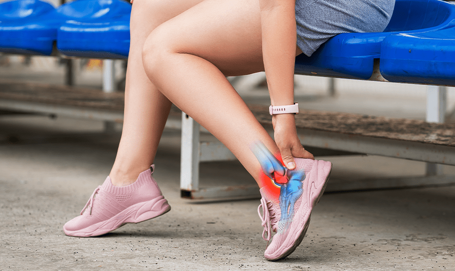
The Achilles tendon is the largest and strongest tendon in the human body. It is formed by the union of the gastrocnemius and soleus muscles (the calf) and attaches to the calcaneus (heel bone). Despite its strength, it is subjected to forces up to several times a person’s body weight during running or jumping.

Achilles injuries occur when the demand placed on the tendon exceeds its structural capacity. This can happen through chronic wear-and-tear (degeneration) or a sudden, explosive force that tears the fibres. A unique characteristic of the Achilles tendon is a “watershed zone”—an area about 2 to 6 cm above the heel attachment—that has a relatively limited blood supply. This zone is where most injuries and ruptures occur, as reduced blood flow can slow the natural repair process.

Types of Achilles Tendon Injuries

Achilles Tendinitis (Acute Inflammation)

Tendinitis involves inflammation of the tendon sheath or the tendon itself. It is usually an overuse injury seen in individuals who have recently increased their training intensity. It is characterised by pain and swelling that often improve with rest but recur with activity.

Achilles Tendinosis (Chronic Degeneration)

Unlike tendinitis, tendinosis is a chronic condition where the tendon’s collagen begins to break down at a microscopic level. The tendon may become thickened and develop small nodules. This condition is often painless in its early stages but can weaken the tendon over time, increasing the risk of a rupture.

Partial Achilles Tendon Rupture

A partial rupture occurs when some of the tendon fibres are torn, but the overall continuity of the tendon remains intact. These can be particularly deceptive because the patient can often still walk, leading to a delay in seeking medical attention. If ignored, a partial tear can easily progress to a complete rupture.

Complete Achilles Tendon Rupture

This is a total separation of the tendon. It most frequently occurs during “stop-and-start” sports. When a rupture occurs, the mechanical link between the calf and the foot is severed, making it impossible to perform movements such as standing on tiptoe or pushing off for a stride.

Acute vs. Chronic Ruptures

An acute rupture is identified shortly after the injury occurs. A chronic rupture is one that has gone undiagnosed for several weeks. In chronic cases, the torn ends of the tendon may have retracted (pulled apart), and scar tissue may have filled the gap, often requiring more complex surgical reconstruction.

Causes & Risk Factors

Direct Mechanisms of Injury

  • Sudden Acceleration: Pushing off forcefully to sprint or jump.
  • Abrupt Direction Changes: Common in court sports like tennis or badminton.
  • Forced Dorsiflexion: A sudden, unexpected trip or fall that forces the foot upward.

Predisposing Risk Factors

  • Age and Gender: Men aged 30 to 50 are most frequently affected, as tendon elasticity naturally decreases with age.
  • “Weekend Warrior” Pattern: Intense exercise on weekends following a sedentary work week.
  • Anatomical Factors: Flat feet (overpronation), high arches, or excessively tight calf muscles can alter the load on the tendon.
  • Medications: The use of certain antibiotics (fluoroquinolones) or corticosteroid injections near the tendon has been associated with weakened tendon structure.
  • Footwear and Surface: Training in worn-out shoes or on hard, unforgiving surfaces like concrete.

Signs & Symptoms

Symptoms of Overuse (Tendinitis/Tendinosis)

  • Stiffness in the morning that gradually loosens with movement.
  • Pain along the back of the leg or heel that worsens after exercise.
  • A visible “bump” or thickening on the tendon.

Symptoms of a Rupture

  • The “Pop”: Many patients report hearing a loud snap or pop at the moment of injury.
  • The “Kick” Sensation: A common description is feeling as though someone has kicked them in the back of the leg, even when no one is there.
  • Gap in the Tendon: A palpable indentation or “soft spot” may be felt a few centimetres above the heel bone.
  • Functional Loss: An immediate inability to push off the foot or stand on the toes of the affected leg.
  • Swelling and Bruising: Rapid onset of swelling around the ankle, followed by bruising that may travel down toward the toes.

Diagnosis & Testing Methods

An orthopaedic evaluation is required to confirm the extent of the injury.

  • The Thompson Test (Calf Squeeze Test): The Thompson test is a commonly used clinical method to check the Achilles tendon. With the patient lying face down, the clinician gently squeezes the calf muscle. In a healthy leg, this causes the foot to point downward. If the Achilles tendon is ruptured, the foot shows little or no movement.
  • Palpation: The surgeon will feel along the tendon for a “gap” or areas of intense tenderness.
  • Ultrasound: This is a dynamic imaging tool that allows the surgeon to see the tendon fibres as the ankle moves. It can help distinguish between a severe partial tear and a complete rupture.
  • MRI (Magnetic Resonance Imaging): MRI provides the most detailed view of the soft tissues. It is used to evaluate the quality of the tendon fibres and determine how far the torn ends have retracted, which is vital for surgical planning.
  • X-rays: While X-rays do not show the tendon, they are used to rule out fractures of the heel bone (calcaneus).

Treatment Options Overview

The choice between surgical and non-surgical management is based on the patient’s age, activity level, and the specific nature of the tear.

Conservative (Non-Surgical) Management

This approach focuses on allowing the tendon to heal naturally.


  • Immobilisation: The foot is placed in a cast or a specialised walking boot, usually with the toes pointed downward (plantar flexion) to bring the torn ends together.
  • Functional Rehabilitation: Modern protocols involve early, protected weight-bearing to encourage the tendon fibres to align correctly as they heal.
  • Suitability: This may be considered for patients who are less active or those with medical conditions that make surgery higher risk.

Surgical Repair

Surgery is often recommended for active individuals and athletes to restore the tendon’s length and tension.


  • Open Repair: A traditional incision is made to allow the surgeon to directly suture the torn ends of the tendon together.
  • Minimally Invasive Repair: Using smaller incisions and specialised instruments, the surgeon can repair the tendon with less disruption to the overlying skin. This may help reduce the risk of wound complications.
  • Tendon Transfers: In chronic or severe cases, the surgeon may move a nearby tendon (such as the tendon that moves the big toe) to reinforce the repaired Achilles.

Biological Augmentation and Injections

  • Platelet-Rich Plasma (PRP): This involves injecting concentrated platelets from the patient’s own blood into the injury site. The growth factors in the platelets may support the healing of the tendon tissue.
  • Shockwave Therapy (ESWT): For chronic tendinosis, shockwave therapy can be used to stimulate blood flow and trigger a healing response in the degenerated tissue.

Wondering which treatment approach may be suitable for your recovery?

A consultation can help you understand the options tailored to your injury.

The Recovery Process: What to Expect

Recovery from an Achilles rupture is a marathon, not a sprint. Adhering to a structured rehabilitation protocol is essential for a successful outcome.

Phase 1: Protection (Weeks 0–6)

The primary goal is to protect the repair (whether surgical or natural). Patients are typically in a boot with “wedges” that keep the heel elevated. Weight-bearing is gradually introduced according to the surgeon’s instructions.

Phase 2: Early Mobilisation (Weeks 6–12)

The heel wedges are removed one by one, bringing the foot back to a neutral position. Physiotherapy focuses on restoring the range of motion of the ankle and beginning very light strengthening exercises.

Phase 3: Strengthening (Months 3–6)

Once the tendon has healed sufficiently, the focus shifts to building calf strength. This often involves “eccentric” exercises, where the muscle is strengthened as it lengthens.

Phase 4: Return to Sport (Months 6–12)

Patients begin straight-line jogging before progressing to agility drills, jumping, and eventually full sports participation. Final clearance for contact sports usually requires the patient to demonstrate near-equal strength between the injured and healthy leg.

Complications

Ignoring an Achilles injury can lead to permanent functional deficits.

  • Elongated Tendon: If a rupture heals with too much scar tissue in the gap, the tendon becomes “too long.” This results in a permanent loss of push-off power, meaning the patient may never be able to run or jump effectively again.
  • Muscle Atrophy: The calf muscle can shrink and weaken significantly (atrophy) if the mechanical link to the heel is not restored.
  • Chronic Pain: Ongoing inflammation and scar tissue can lead to persistent discomfort during daily walking.

Prevention and Tendon Health

While not all injuries can be prevented, certain strategies can reduce the risk:

  • Gradual Progression: Avoid increasing your exercise intensity or duration.
  • Proper Footwear: Ensure your shoes provide adequate heel support and are replaced once the cushioning begins to fail.
  • Calf Flexibility: Regular, gentle stretching of the calf muscles can reduce the tension on the Achilles tendon.
  • Warm-ups: Always perform dynamic warm-ups (like leg swings or light jogging) before engaging in explosive sports.
Achilles Tendon Injury

Frequently Asked Questions

Can I walk on a ruptured Achilles tendon?

Some people can walk flat-footed by using other muscles, but they will not be able to “push off” or stand on their tiptoes. Walking on a rupture without a boot can cause the torn ends to pull further apart, complicating future treatment.

How long is the hospital stay for surgery?

Many Achilles repairs are performed as day-case surgeries, meaning the patient can return home the same day. Some may require an overnight stay for pain management.

Will I need to wear a cast?

Most modern protocols have moved away from long-term plaster casts in favour of functional walking boots, which allow for earlier movement and better hygiene.

When can I drive again?

This depends on which leg is injured and whether you drive an automatic or manual vehicle. If the right leg is injured, you typically cannot drive until you are out of the boot and have regained sufficient braking strength, which usually takes about 8 to 12 weeks.

Conclusion

An Achilles tendon injury is a significant challenge, but with the right clinical approach, recovery is achievable. Whether managed through surgery or conservative therapy, the focus remains on restoring the strength and flexibility of this vital tendon. In Singapore, patients have access to advanced orthopaedic techniques and specialised sports physiotherapy to support their journey back to health.

Early assessment is key. If you suspect a tendon injury, seeking professional advice can help ensure that you receive the most appropriate care for your specific needs.

Ready to Take the Next Step?

If you’re experiencing heel pain or suspect a tendon rupture, a consultation with an orthopaedic surgeon can provide a clear diagnosis and a structured recovery plan.

Dr Zackary Chua 1

Dr Zackary Chua (蔡克祥医生)

MBBS (AUS)|MMed (Singapore)|MRCS (Glasgow)|FRCSEd (Orth)

A senior consultant orthopaedic surgeon with dual specialisations in Foot & Ankle Surgery and Paediatric Orthopaedics. Backed by his extensive years of training and experience, Dr Chua brings the necessary knowledge and skills to ensure a smooth recovery journey.

Rooted firmly by two fundamental philosophies: “When one’s feet hurt, one hurts all over” and “Children are not little adults”, Dr Chua’s approach to patient care reflects his compassionate care for adults and children.

  • Singhealth Quality Excellence Award – Silver (2011)
  • Service From the Heart Award (2021)
  • Singapore Health Quality Service Award – Gold (2023)
  • NUS Medicine Dean’s Award for Teaching Excellence (2023)

His active participation in the medical community extends to mentoring, research supervision, and contributions to key professional forums and symposiums, both locally and internationally.

Your Treatment Roadmap

Registration

Our friendly clinical staff will assist you with your registration, ensuring an efficient and hassle-free process for you.

Specialist Consultation

During your consultation, we will evaluate your medical history and the pain you are experiencing. You may also ask questions about your condition.

Diagnosis Of Your Condition

We will likely perform some form of diagnostic imaging (e.g. X-Rays, MRI) to assess your condition accurately.

individualised Treatment Plan

After imaging, we will review the results of your scans thoroughly, and advise an individualised treatment plan for you.

Follow-Up Visits

As you go through your treatment plan, we will follow up with you every step of the way, ensuring that you receive the dedicated care you deserve.

Billing & Insurance

Integrated Shield Plans & Corporate Insurance

For Singaporeans, Singapore Permanent Residents and Foreigners.
Please speak to our friendly clinic staff about using your insurance plans.

Make An Enquiry

Got a Question? Fill up the form and we will get back to you shortly.

    Full Name*

    Email Address*

    Phone Number*

    Your Message*

    For Faster Response, Call us!

    +65‎ 6956‎ 6588


    Our Locations

    Mount Elizabeth Novena Specialist Centre
    38 Irrawaddy Road, #05-36, Singapore 329563

    +65‎ 6956‎ 6588 (24 hours) (Calls / Messages)
    +65 8877 3318 (24 hours) (WhatsApp)
    Monday – Friday: 9am – 5pm
    Saturday: 9am – 12:30pm
    Sunday & PH: Closed

    Parkway East Medical Centre
    319 Joo Chiat Place, #03-06, Singapore 427989

    +65‎ 6956‎ 6588 (Calls / Messages)
    +65 8877 3318 (WhatsApp)
    Monday – Friday: 9am – 5pm
    Saturday: 9am – 12:30pm
    Sunday & PH: Closed
    (Parkway East Clinic is on appointment basis)